
The Hidden Cost of Poor Imaging Quality in Neurodegenerative Disease Clinical Trials
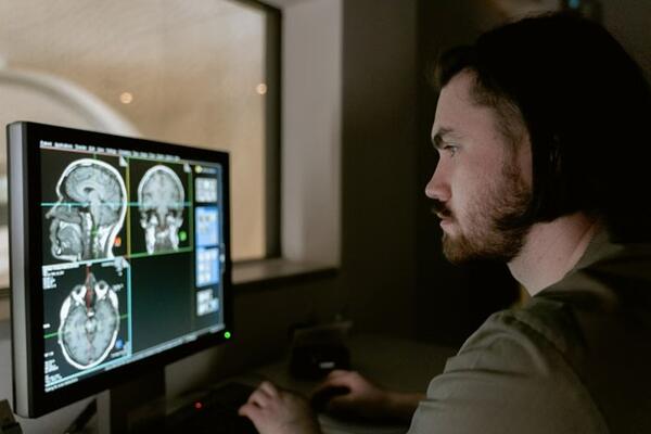
Imaging data sits at the foundation of most neurodegenerative disease clinical trials, and when that foundation is unstable, the costs compound quickly. MRI and PET scans are rarely just diagnostic tools in these studies; they serve as primary endpoints, tracking subtle changes in brain structure and metabolic activity that no other measure can capture with the same precision.
When imaging data is compromised by variability, the downstream effects move well beyond the scanner room. Unusable scans require repeats, protocol deviations trigger manual review, and inconsistent data forces statistical teams to increase sample sizes just to recover the power that poor image quality eroded. Each of these problems carries a direct operational cost, but they also extend timelines in ways that affect site contracts, patient retention, and regulatory submissions.
What makes imaging errors particularly difficult to manage in neurodegenerative trials is that the signal being measured is often small to begin with. Studies tracking early Alzheimer's progression or subtle atrophy patterns leave little margin for noise. For participants considering why seniors should consider joining clinical trials, the stakes are just as real, because data quality shapes whether a study ultimately delivers answers.
Where Poor Imaging Quality Gets Expensive Fast
Imaging biomarkers are the technical category at the center of trial cost exposure in neurodegenerative studies. When imaging biomarkers are used for enrollment, disease progression tracking, or endpoint assessment, any acquisition inconsistency multiplies downstream cost across the study. The financial exposure is not limited to the imaging operation itself; it spreads into statistical planning, site management, and regulatory timelines.
Unusable scans, repeat imaging events, and protocol deviations each generate immediate operational costs. Beyond those direct expenses, data noise forces sponsors to increase sample sizes to recover statistical power, which extends enrollment windows and delays milestones. The problem is both scientific and financial, and treating it as a narrow technical issue consistently underestimates its true scope.
For participants considering why seniors should consider joining clinical trials, the integrity of imaging data is equally consequential, since the quality of that data shapes whether a study ultimately delivers answers that reach patients.
Why Neurodegenerative Trials Are Uniquely Exposed
Neurodegenerative trials operate in a space where the biology itself creates measurement challenges that most other therapeutic areas do not face. The changes being tracked, including gradual brain atrophy, shifting amyloid burden, and slow dopaminergic decline, unfold over months or years, and the differences between treatment and placebo arms can be remarkably small. That biological reality makes imaging quality failures disproportionately damaging compared to other trial types.
MRI and PET Carry Different Failure Risks
Structural MRI and volumetric MRI are sensitive to acquisition variability in distinct ways. Field strength inconsistencies, head positioning shifts, and gradient calibration drift can all introduce measurement error that mimics or masks genuine tissue loss. In Alzheimer's disease and Parkinson's disease trials, where brain atrophy rates over 12 months may measure in fractions of a millimeter, even minor hardware inconsistencies become statistically meaningful.
PET imaging introduces a separate category of risk. Tracer variability, scanner sensitivity drift, and reconstruction parameter differences across sites affect quantitative imaging outputs in ways that structural sequences do not. When amyloid burden or metabolic activity is the primary endpoint, those inconsistencies directly distort the treatment signal being evaluated.
Small Signal Changes Raise the Price of Noise
Longitudinal clinical trial imaging amplifies these problems because each site visit adds another opportunity for protocol drift to accumulate. Peer-reviewed research has documented how multi-site inconsistency in neuroimaging studies widens variance and reduces statistical power, effectively requiring larger samples to detect the same effect.
Disease progression in neurodegenerative conditions rarely produces dramatic imaging shifts, which means noise does not just add error. It can obscure a real treatment effect entirely, producing false negatives that bury genuinely effective therapies.
How Quality Failures Spread Through a Trial
Understanding where quality failures originate at the site level makes it easier to appreciate how quickly they escalate into trial-wide consequences. The causal chain runs from individual scan rooms to sponsor budgets, and each link in that chain carries its own cost.
At the Site Level, Errors Start With Acquisition
Most quality failures in clinical trial imaging begin at the point of acquisition, where protocol compliance is hardest to enforce consistently across sites. Wrong pulse sequences, timing deviations from the approved protocol, uncorrected motion artifacts, and inconsistent reconstruction settings are among the most common sources of degraded data.
Each of these errors creates an immediate operational consequence. A scan acquired with incorrect parameters may need to be repeated, a protocol deviation requires query resolution between the site and the imaging core, and data that cannot be salvaged is flagged for exclusion. None of these outcomes are trivial; repeat imaging consumes site resources and reimbursement budgets, while query cycles introduce administrative delays that accumulate across hundreds of visits.
For MRI and PET studies tracking slow neurodegeneration, a single excluded timepoint can break the longitudinal data chain for that subject, reducing the integrity of individual trajectories before the study-level effects are even considered.
At the Study Level, Variability Reshapes Outcomes
When imaging errors are isolated, the impact is manageable. When they occur systematically across sites, however, the consequences scale to the trial level. Missing or noisy scans reduce the evaluable population available for endpoint analysis, shrinking the set that statistical models depend on.
Reduced endpoint analysis sets have a direct relationship with statistical power. As usable data decreases, sponsors face a choice between accepting reduced confidence in results or extending enrollment to recover the sample size that standardization failures eroded. Either path carries cost; enrollment extensions delay milestone timelines, stretch site contracts, and push regulatory submissions further out.
Poor imaging quality, in this way, converts a scan-room problem into a budget and timeline event that reaches well beyond the imaging operation itself.
The Regulatory Risk Is Larger Than It Looks
The operational costs described in the previous sections are significant, but they represent only part of the exposure. When imaging quality problems persist into the submission package, they create a separate category of risk that unfolds during FDA review rather than during the trial itself.
Endpoint inconsistency is one of the most consequential issues a submission can carry. When imaging biomarkers show unexplained variability across sites or timepoints, reviewers may question whether the observed treatment effect reflects genuine disease progression changes or measurement artifact. That doubt rarely resolves in a sponsor's favor without additional analysis.
Imaging anomalies in a submitted dataset tend to trigger further scrutiny. Regulators may request sensitivity analyses to test whether results hold after excluding problematic scans, or they may issue clarification requests that pause the review clock while sponsors reconstruct the evidentiary basis for their claims. Each of these responses extends the timeline and introduces uncertainty that was avoidable.
The deeper issue is that a compromised quantitative imaging dataset weakens confidence in the treatment effect claim at precisely the moment that confidence needs to be strongest. In neurodegenerative diseases, where endpoint sensitivity is already limited by the biology, reviewers have limited tolerance for noise that further obscures the signal. This is why imaging quality assurance belongs inside regulatory strategy from the outset, rather than being treated as a separate operational matter that gets addressed only after problems surface during review.
What Reduces Imaging Risk Before It Compounds
Addressing imaging risk early is consistently less expensive than correcting problems after they have propagated through a study. The sections above make clear how quickly site-level errors become trial-level consequences, which is why the most effective mitigation strategies focus on prevention rather than remediation.
Site Qualification and Central Review Matter Most
Prevention consistently costs less than remediation in clinical trial imaging, and the evidence from large-scale efforts like ADNI reinforces that standardization built into the study infrastructure, rather than applied as a correction after problems emerge, is what keeps imaging data usable.
Site qualification is where that infrastructure begins. Confirming that MRI and PET scanners meet protocol specifications before enrollment opens, harmonizing acquisition parameters across locations, and delivering protocol compliance training to imaging staff reduces the variability that compounds over time in multi-site studies.
Blinded Independent Central Review adds a second layer of protection by evaluating imaging data outside the site environment, catching acquisition errors, read inconsistencies, and protocol deviations before they reach analysis lock. Centralized quality control creates feedback loops that allow corrective guidance to reach sites while the study is still running, rather than after the evaluable population has already been reduced. For families and clinicians thinking about memory care planning for Alzheimer's and dementia, the integrity of trial data directly shapes what treatments eventually reach patients.
FAQs
How Does Poor Imaging Quality Affect Neurodegenerative Disease Clinical Trials?
It reduces evaluable data, forces repeat scans, and can require larger sample sizes to recover lost statistical power, extending timelines and increasing costs.
Why Are MRI and PET Scans Especially Sensitive in These Trials?
MRI detects subtle structural changes like atrophy, while PET measures metabolic activity and amyloid burden. Both are vulnerable to acquisition variability that distorts small treatment signals.
What Is Blinded Independent Central Review in Imaging Trials?
Blinded Independent Central Review evaluates imaging data independently from trial sites, catching protocol deviations and read inconsistencies before they compromise the final analysis dataset.
Can Imaging Quality Problems Delay FDA Review?
Yes. Unexplained variability in imaging biomarkers can prompt regulators to request additional analyses or clarifications, pausing the review clock and extending the overall submission timeline.
Imaging Quality Is a Budget Decision
Poor imaging quality in clinical trial imaging does not create a single large failure; it creates a sequence of smaller ones that each carry a cost. Repeat scans, query cycles, excluded subjects, and reduced statistical power accumulate into timeline delays and budget overruns that often exceed what prevention would have required.
Standardization and central oversight are not procedural formalities. For neurodegenerative diseases, where imaging biomarkers carry much of the evidentiary weight, they are financially rational choices that protect both the science and the submission.
The decision about imaging quality is ultimately a budget decision. Sponsors who treat it as one from the outset give FDA reviewers cleaner data and give themselves a more defensible path to approval.